Professor David Knecht: ‘Breadcrumb trail’ of fatty acid drives aggressive spread of melanoma.

October 16, 2014

Melanoma is an unusually aggressive cancer which spreads or metastasizes very quickly early in the tumor development. The driver of this spread away from the primary tumor has not been well understood. Now results of a new study have shown that melanoma cells follow a ‘breadcrumb trail’ of a fatty serum component called lysophosphatidic acid (LPA).

For more, go to:

Andrew J. Muinonen-Martin, Olivia Susanto, Qifeng Zhang, Elizabeth Smethurst, William J. Faller, Douwe M. Veltman, Gabriela Kalna, Colin Lindsay, Dorothy C. Bennett, Owen J. Sansom, Robert Herd, Robert Jones, Laura M. Machesky, Michael J. O. Wakelam, David A. Knecht, Robert H. Insall (2014) Melanoma Cells Break Down LPA to Establish Local Gradients That Drive Chemotactic Dispersal. PLoS Biology. DOI: 10.1371/journal.pbio.1001966

The Guardian article.


MCB joins in the welcome for our colleagues at the new Jackson Laboratory Center for Genomic Medicine

October 9, 2014

MCB is developing many connections with the new Jackson Laboratory for Genomic Medicine that has just opened on the Farmington Campus.  A number of MCB faculty have already submitted grant proposals with JaxGM faculty, there are internship agreements with the Applied Genomics and Microbial Systems Analysis professional science master’s programs in MCB, as well as several other initiatives under discussion.  MCB welcomes JaxGM to the Connecticut biomedical research community, and looks forward to a strong relationship with our new colleagues.

Dr. Eric May and Graduate Student Jason Pattis to run simulations on world’s fastest computer for molecular dynamics.

October 6, 2014

Through a competitive application process run by the Pittsburgh Supercomputing Center, Dr. May and Pattis have received an allocation on the Anton supercomputer. Anton is a specialized supercomputer designed by D.E. Shaw Research for the sole purpose of performing molecular dynamics simulations of biomolecules. May and Pattis will use their 50,000 node-hour allocation to compute microsecond dynamics of a protein from the deadly Lassa virus.

MCB Undergrads Making the News!

October 2, 2014

Click to view the NBC video.

UConn students, CT Children’s medical researchers and children and their families are partnering in a novel collaboration and changing the face of medical care for kids in Connecticut. The UCONN students in the Undergraduate Research Associate Program (URAP) in the Department of Molecular & Cell Biology, work closely with physicians in the Department of Pediatrics at Connecticut Children’s Medical Center (CT Children’s).  Bridging the students’ basic science training to clinical problems in pediatric care and diet has improved treatments for children at CT Children’s.  The work from these students and their physician mentors has resulted in several peer reviewed clinical publications and presentations at national meetings.

In a development with enormous potential, the URAP program is now partnering with Kids and Families Impacting Disease Through Science (KIDS).  Partnering with KIDS will allow this collaborative research to reach its full potential for children.  KIDS is an advisory group of children, adolescents and families.  They focus on understanding, communicating and improving medicine, research and innovation for children; thus perfect partners for the URAP program.  (KIDS is a collaboration between the American Academy of Pediatrics (AAP) Section on Advances in Therapeutics and Technology (SOATT), local AAP Chapters, children’s hospitals, local schools and other partners.)  This new partnership brings together diverse and important stakeholders in the future of health care for all our children.

This hugely beneficial experience for UCONN students has matured into an incredibly effective program that simultaneously produces, through research, critical advantages in health care delivery for CT Children’s pediatricians and children and their families.

MCB 3189 Instructors: Arlene Albert and Sharon Smith.

The 2014 Khairallah Symposium: Tuesday, October 7, 2014. Invited Seminar: “Electron Cryomicroscopy of Rotary ATPases” by Dr. John Rubinstein.

September 29, 2014

Since 1997, the Dr. Edward A. Khairallah Fund for Scholarship has supported ongoing symposia and support for graduate students pursuing degrees in biochemistry, cell biology or toxicology. This year’s Symposium will pay tribute to his late wife, Dr. Lamia H. Khairallah, and her enduring contributions to teaching and research at the University of Connecticut. It also marks the renaming of the Fund to the Dr. Edward A. and Dr. Lamia H. Khairallah Fund for Scholarship.

A catered reception will be held at 3:30 pm in Room 201 of the Biology/Physics Building. This event will feature a presentation by Dr. Steven Cohen and members of the Khairallah Family.

The Khairallah Memorial Seminar will be held at 4:00 pm in Room 130 of the Biology/Physics Building. This talk, entitled “Electron Cryomicroscopy of Rotary ATPases” will be presented by Dr. John Rubinstein from the University of Toronto. To schedule time to meet with Dr. Rubinstein, please contact the host, Dr. Nathan Alder (nathan.alder@uconn.edu).

Professor Burkhard’s in UConn Today

September 18, 2014

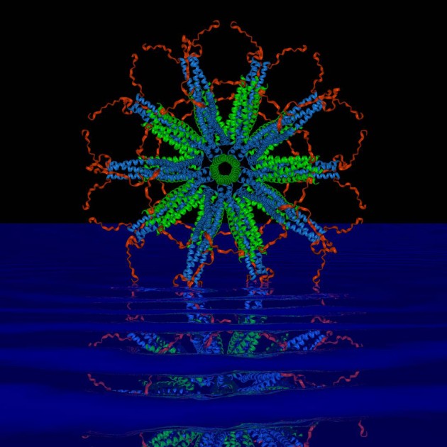
A computer image of a self-assembling protein nanoparticle that relies on rigid protein structures called 'coiled coils' (blue and green in the image) to create a stable framework upon which scientists can attach malaria parasite antigens. (Image courtesy of Peter Burkhard)

A self-assembling nanoparticle designed by a UConn professor is the key component of a potent new malaria vaccine that is showing promise in early tests.

For years, scientists trying to develop a malaria vaccine have been stymied by the malaria parasite’s ability to transform itself and “hide” in the liver and red blood cells of an infected person to avoid detection by the immune system.

But a novel protein nanoparticle developed by Peter Burkhard, a professor in the Department of Molecular & Cell Biology, in collaboration with David Lanar, an infectious disease specialist with the Walter Reed Army Institute of Research, has shown to be effective at getting the immune system to attack the most lethal species of malaria parasite, Plasmodium falciparum, after it enters the body and before it has a chance to hide and aggressively spread.

The key to the vaccine’s success lies in the nanoparticle’s perfect icosahedral symmetry (think of the pattern on a soccer ball) and ability to carry on its surface up to 60 copies of the parasite’s protein. The proteins are arranged in a dense, carefully constructed cluster that the immune system perceives as a threat, prompting it to release large amounts of antibodies that can attack and kill the parasite.

In tests with mice, the vaccine was 90-100 percent effective in eradicating the Plasmodium falciparum parasite and maintaining long-term immunity over 15 months. That success rate is considerably higher than the reported success rate for RTS,S, the world’s most advanced malaria vaccine candidate currently undergoing phase 3 clinical trials, which is the last stage of testing before licensing.

Peter Burkhard, professor of molecular and cell biology, with a computer image of the protein nanoparticle he designed. (Peter Morenus/UConn Photo)

“Both vaccines are similar, it’s just that the density of the RTS,S protein displays is much lower than ours,” says Burkhard. “The homogeneity of our vaccine is much higher, which produces a stronger immune system response. That is why we are confident that ours will be an improvement.

“Every single protein chain that forms our particle displays one of the pathogen’s protein molecules that are recognized by the immune system,” adds Burkhard, an expert in structural biology affiliated with UConn’s Institute of Materials Science. “With RTS,S, only about 14 percent of the vaccine’s protein is from the malaria parasite. We are able to achieve our high density because of the design of the nanoparticle, which we control.”

The research was published in Malaria Journal in 2013.

The search for a malaria vaccine is one of the most important research projects in global public health. The disease is commonly transported through the bites of nighttime mosquitoes. Those infected suffer from severe fevers, chills, and a flu-like illness. In severe cases, malaria causes seizures, severe anemia, respiratory distress, and kidney failure. Each year, more than 200 million cases of malaria are reported worldwide. The World Health Organization estimated that 627,000 people died from malaria in 2012, many of them children living in sub-Saharan Africa.

It took the researchers more than 10 years to finalize the precise assembly of the nanoparticle as the critical carrier of the vaccine and find the right parts of the malaria protein to trigger an effective immune response. The research was further complicated by the fact that the malaria parasite that impacts mice used in lab tests is structurally different from the one infecting humans.

The scientists used a creative approach to get around the problem.
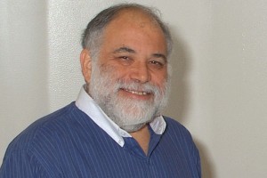
Infectious disease specialist David Lanar of the Walter Reed Army Institute of Research, who is collaborating with UConn professor Peter Burkhard in pursuing a new malaria vaccine. (Photo courtesy of David Lanar)

“Testing the vaccine’s efficacy was difficult because the parasite that causes malaria in humans only grows in humans,” Lanar says. “But we developed a little trick. We took a mouse malaria parasite and put in its DNA a piece of DNA from the human malaria parasite that we wanted our vaccine to attack. That allowed us to conduct inexpensive mouse studies to test the vaccine before going to expensive human trials.”

The pair’s research has been supported by a $2 million grant from the National Institutes of Health and $2 million from the U.S. Military Infectious Disease Research Program. A request for an additional $7 million in funding from the U.S. Army to conduct the next phase of vaccine development, including manufacturing and human trials, is pending.

“We are on schedule to manufacture the vaccine for human use early next year,” says Lanar. “It will take about six months to finish quality control and toxicology studies on the final product and get permission from the FDA to do human trials.”

Lanar says the team hopes to begin early testing in humans in 2016 and, if the results are promising, field trials in malaria endemic areas will follow in 2017. The required field trial testing could last five years or more before the vaccine is available for licensure and public use, Lanar says.

Martin Edlund, CEO of Malaria No More, a New York-based nonprofit focused on fighting deaths from malaria, says, “This research presents a promising new approach to developing a malaria vaccine. Innovative work such as what’s being done at the University of Connecticut puts us closer than we’ve ever been to ending one of the world’s oldest, costliest, and deadliest diseases.”

A Switzerland-based company, Alpha-O-Peptides, founded by Burkhard, holds the patent on the self-assembling nanoparticle used in the malaria vaccine. Burkhard is also exploring other potential uses for the nanoparticle, including a vaccine that will fight animal flu and one that will help people with nicotine addiction. Professor Mazhar Khan from UConn’s Department of Pathobiology is collaborating with Burkhard on the animal flu vaccine.

article by Colin Poitras
Dr. Barbara Mellone was awarded a $510,000 grant from the NSF, “Characterization of a novel, evolutionarily distinct chaperone for centromeric histone H3.”

September 2, 2014

Abstract:

This research will take an evolutionary approach to understand how centromeres assemble. Centromeres are essential elements of chromosomes that are crucial for proper transmission of genetic information from cell to cell during cell division. When centromeres fail to assemble correctly, the result can be an imbalance in chromosome number whereby cells have too many or too few chromosomes. In animals, such an imbalance can lead to visible developmental defects, which in turn can lead to birth defects or death. This study aims to impact our understanding of the conserved aspects of centromere assembly, and hence aspects that are crucial for preventing an imbalance in chromosome number and catastrophic developmental defects. In addition, this research will have a strong educational impact by providing opportunities for undergraduate and graduate students to engage directly in research and by developing a new hands-on summer laboratory workshop for high school teachers to enable them to learn about chromosome biology and take the lessons learned back to their high school classrooms.

In eukaryotes, centromeres assemble by binding special centromere-specific histone H3 proteins, which are escorted to the correct location on the chromosome by chaperone proteins. Recent results have revealed structural and sequence variation in these chaperones, which suggests that during evolution, multiple mechanisms may have arisen for assembling centromeres. This work will focus on analysis of the chaperone proteins using evolutionary, structural and functional studies. Comparisons will be made among a newly discovered chaperone from Drosophila and two well-conserved chaperones from yeast and mouse. The outcomes of the work will provide unique mechanistic and evolutionary insights into the fundamental processes that govern centromere structure and function. The project will also have broad impact from the educational perspective. The research will be carried out by undergraduate and graduate students, many of whom are women, thereby providing opportunities for scientific training, career development and direct participation in the promulgation of scientific findings through publications and conference presentations. In addition, a novel hands-on summer laboratory workshop will be developed for high school teachers (ten per year), focusing on the basic biology of chromosome segregation and cell division and extending to emerging topics such as stem cells and altered genetic states in cancer. The teachers will leave the workshop armed with teaching materials, ideas for lesson plans, and positive research experiences through their direct involvement in experimentation. By enabling the teachers to take what they learn back to their classrooms, the workshops will have a multiplier effect on the scientific education of scores of pre-college level students.

J. Peter Gogarten ​ (MCB)​ , R. Thane Papke ​ ​ (MCB), and Uri Gophna (Tel Aviv University, Israel) were awarded a grant by the Unites States – Israel Binational Science Foundation to study “How selfish elements impact and reflect speciation and recombination in archaea”

August 21, 2014

ABSTRACT
A major challenge in evolutionary biology is to understand the processes of
divergence and diversification in microorganisms, in which genes can often be
exchanged laterally and not just from parent to offspring. The proposed research will
test how a special class of selfish elements, termed homing endonucleases (HEs),
affects the genetic barriers that can form distinct groups of microorganisms, often
labeled as ecotypes or species, and use HEs to estimate how much lateral gene
exchange exists within a natural population. We have established the experimental
framework required to attain these goals in halophilic Archaea using several
complementary approaches: theoretic-computational (UConn), genetic (TAU),
genomic (UConn) and ecological (TAU). We will test how the presence of HEs can
affect gene exchange within and between groups, and investigate the spread of HEs
and its effects on recombination in an ecological context using a collection of
environmental isolates. We will perform genetic experiments and use the results
derived from them to refine computational models that describe HE dynamics. We
will then generalize these findings and generate models for the effects of HEs on
microorganism population genetics, testing them using computer simulations. The
proposed research will shed new light on the role that mobile parasitic elements play
in shaping microbial evolution.

John Malone was invited to particpate in the New Horizons in Science symposium.

June 24, 2014

The symposium is conducted by the Royal Society of Canada, The Mexican Academy of Sciences, and the US National Academy of Science. The science academies of Canada, Mexico and the United States are working towards broader science cooperation between our countries by building relationships among future science leaders. As a first step, this joint symposium focuses on young to mid career scientists working in astrophysics, biotechnology, green chemistry, marine science and the science of hazards and disasters

For more information about the symposium, click here.