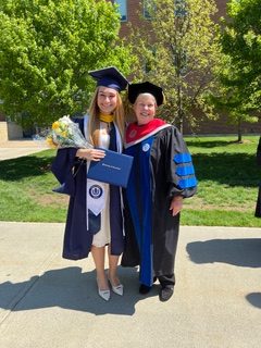
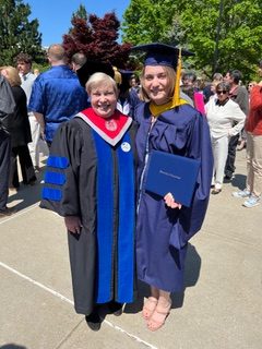
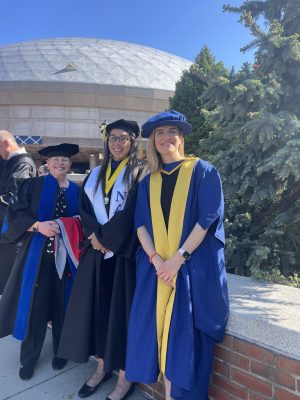
June 11, 2024

A Cool Spin on Large Biomolecular Complexes

Our newest MCB colleague Professor Dylan Murray received his undergraduate training at SUNY-Plattsburgh in Physics. After graduating, he spent three years in an X-ray crystallography laboratory at the University of Vermont studying surface charge density in the enzyme lysozyme. His exposure to biological research convinced him to pursue doctoral work developing solid-state NMR (ssNMR) methods to study membrane protein drug targets involved in tuberculosis at the National High Magnetic Field Laboratory of Florida State University. His doctoral work culminated in a Ph.D. thesis entitled “The structure of a three-helix membrane protein in a lipid bilayer”. Following this, Dr. Murray was scouted by the NIH where he did postdoctoral work in a ssNMR laboratory famous for work on amyloids, learning to use these methods to study disordered proteins that form membrane-less organelles. Professor Murray joins us from University of California, Davis where he used ssNMR to research diverse topics including RNA granules, cell cytoskeleton proteins, and plant cell walls. Professor Murray brings new strengths in ssNMR to complement our existing structural biology and biophysics programs. We were fortunate to simultaneously recruit a second ssNMR spectroscopist, Joana Paulino to our sister institution UConn Health.

The past half-century has seen an explosion in our knowledge of molecular structure. This data informs how molecules function and how they can be targeted by drugs in cases of dysfunction. The Protein Data Bank (PDB) currently holds over 200,000 experimental structures of biological macromolecules and over 1,000,000 AlphaFold predictions generated using AI algorithms, that predict protein models using features learned from the ever-growing database of known experimental structures. The PDB repository of molecular structures has been invaluable in combatting the COVID-19 pandemic, in drug and protein design efforts, and will continue to be a tremendous resource in efforts to understand biological mechanism at a quantitative level, beyond polymer sequencing and vague homology-based functional inferences.

The primary experimental methods to determine macromolecular structures experimentally have been X-ray crystallography, cryo-electron microscopy (cryo-EM) and solution nuclear magnetic resonance (NMR). Each method has its strengths and weaknesses. X-ray crystallography requires molecules to be coaxed into ordered crystalline arrays, which is not always achievable. Cryo-EM uses electron microscopes to directly image molecules but the signal from individual molecules is too weak, so that averaging over hundreds of thousands to millions of molecules is required to achieve the resolution needed for atomic detail. For this type of averaging to work, the molecules must be sufficiently large and identical, as disorder or heterogeneity will smear out the signal. Solution NMR can be used to study biological molecules in their natural aqueous environment, whether ordered or disordered, but the technique has a frustrating size limit that makes it extremely difficult to study molecules or complexes much larger than 50 kDa. Larger molecules give broader NMR signals because they tumble more slowly in solution. The most recent addition to the arsenal of methods to shed light on biomolecular structure is ssNMR that has currently contributed some 175 structures in the PDB. Under ordinary circumstances, ssNMR is plagued by extremely broad lines resulting in poor signal-to-noise in experiments, since in a gel-like solid state molecules do not tumble at all. However, practitioners of the technique have ingeniously optimized ever-faster methods to mechanically spin their samples in rotors at the “magic angle” (arctan√2 = 54.7o). Spinning speeds > 100 kHz have recently been achieved, much faster than a dentist’s drill but still considerably slower than small molecules spin in solution (~ 1 GHz). Because spinning in ssNMR is mechanical, there is no physical restriction on the size of the molecules in the sample that can be studied. The ssNMR method has therefore had tremendous success in characterizing some of the largest and most challenging targets in structural biology: large assemblies including viruses, amyloid fibrils involved in neurodegenerative diseases, as well as some more standard globular protein complexes.

Professor Murray’s laboratory at UConn will focus on three research areas. (1) One focus of the laboratory is low complexity proteins in membrane-less organelles. Under conditions of stress or during normal cell function, cells can form structures such as Cajal bodies, nuclear speckles, P-bodies, or RNA processing granules, that segregate their contents using a phase separation process akin to oil and water in salad dressing, but involving intrinsically denatured proteins (IDPs), or IDPs in complexes with RNA. A key feature of the proteins that form membrane-less organelles are IDP regions with repetitive amino acid sequences that allow them to form a meshwork to stabilize the phase-separated liquid droplet complex. However, the structural promiscuity needed for the flexibility of these proteins appears to come at a price — over time, the liquid droplets can age into rigid aggregates that are associated with several protein misfolding diseases such as Huntington’s, Parkinson’s, Alzheimer’s and Lou Gehrig’s disease. The Murray Laboratory is interested in the mechanism of these structural transitions, with an eye to how they might be targeted by therapeutic interventions. (2) Intermediate filaments are a second research focus of the Murray Laboratory. The filaments are multichain assemblies built through helical coiled-coil interactions to form fibers with an average diameter of 10 nm. This distinguishes intermediate filaments from the thin 7 nm actin, and thick 25 nm microtubules fibers in cells. Intermediate filaments are a structural building block for a variety of tissues including hair, skin, nails, and feathers but the Murray laboratory is specifically interested in filaments that provide structural support for glial cells, part of the immune system for the nervous system, and cells that are subject to cancerous transformation. The aim is to understand how the coiled-coil regions together with disordered head and tail regions determine filament assembly and structural properties, and how assembly goes awry due to pathological mutations linked to Alexander’s Disease and how cancerous cells exploit it during metastasis. (3) Plant cell wall structure constitutes a third research focus where the aim is to provide a 3D picture of the intact cell wall and how it changes during the extraction of biofuels and chemical precursors traditionally obtained from the petrochemical industry. In collaboration with plant geneticists, the Murray Laboratory is exploring if mutations in the protein machinery constructing the cell wall can convert these biological materials to rich sources of energy and chemical alternatives.

A ”cool” aspect of the new instrumentation Professor Murray will use at UConn is a cryogenic probe (cryoprobe) that will be one of the first available for ssNMR in the world. Cryoprobes cool the electronic components used to detect the NMR signal (but not the actual sample) to liquid helium temperatures (25 K), achieving a boost in sensitivity by reducing thermal noise in the electronics. The boost in sensitivity allows for faster data collection that opens a window for studying very small amounts of difficult to obtain samples or kinetic experiments to interrogate how samples change over time, for example, to follow processes such as disease-related aggregative misfolding that the Murray Laboratory is investigating.

For the future, Professor Murray sees continuing improvements in ssNMR technology including probes that can achieve faster spinning rates, sensitivity boosts through dynamic nuclear polarization (DNP), and improved sample isotope labeling schemes that can clarify the complex NMR spectra of large macromolecular complexes. Beyond the types of projects currently under study, he envisions his research could lead to the design of self-assembling nanostructures for drug development or contrast agents for bioimaging, and other advances that will facilitate the use of ssNMR in studying molecules within living cells or to follow metabolism in tissues.

Professor Murray is slated to teach Biophysical Chemistry I in Fall 2024. He plans to eventually offer additional advanced NMR and phase separation in biology courses. In terms of his research laboratory, students can expect to receive broad and collaborative training in several areas depending on their strengths, interests, and laboratory needs. Students might work on protein purification, imaging, and assay development or delve more heavily into NMR instrumentation or data analysis and computation. He emphasizes that potential students should not be intimidated by their unfamiliarity with NMR and notes “If you’re excited about the science you will get the training you need!”. Contemporary biology is more about addressing biological problems than techniques. Researchers today are expected to learn and adapt to whatever technique is needed to solve the biological problem. Professor Murray actively fosters this type of learning in his laboratory and integrates it into the training plan for all members of the research team.

MCB is delighted to have a new colleague that brings strengths in ssNMR and macromolecular assemblies — research areas that promise to achieve cool new results for decades to come.


GO:MCB Announces 24-25 Executive Board

June 5, 2024

GO:MCB has announced the results of the 24-25 e-board election.
President: Josette Nammour
Vice President: Katherine Scalise
Secretary: Heather Jamieson
Outreach Coordinator: Shania Kalladanthyil
Diversity Facilitator: Mullein Francis
DEIC Grad  Representatives: Sophia Gosseline and Katherine Fleck

Education in Aging and Geroscience Research Program (EAGR) announces 2024 Scholars

June 3, 2024

EAGR Program Director, Kenneth Campellone is pleased to announce that six undergraduate students have been named as 2024 scholars.

2024 EAGR Scholars
L>R Milad Zameeri, Emily Szydlo, Lizzy Parkerson, RoseMina Notch, Bailey Millis, Kelsey Gorgei

Bailey Millis, Kelsey Gorgei, Emily Szydlo, Elizabeth Parkerson, Milad Zameeri, and RoseMina Notch will participate in an immersive, hands-on Summer research program in Geroscience research labs at UConn and UConn Health.

The UConn Undergraduate Education in Aging and Geroscience Research Program is an education and research initiative sponsored by the National Institute on Aging. (NIA) The program integrates coursework, research, and outreach activities.

Learn more about the EAGR Program


Arcane Archaea

May 20, 2024

The Santiago-Martinez lab uses modern approaches to investigate ancient things.

-by Stacey Hanlon

Cellular life on Earth can be classified into one of three domains: Eukarya (Eukaryota), Bacteria, and Archaea.

Geo Santiago-Martinez
Dr. Geo Santiago-Martínez, Principal Investigator at the UConn Microbial Ecophysiology Laboratory.

Though most of us are familiar with species that fall into the Eukaryota and Bacteria domains, those belonging to the Archaea domain are often ignored despite being found all around us.  From the Greek word meaning “ancient things,” archaea are found in a variety of harsh environments like boiling-hot geysers and salt lakes, but they can also be found in the soil and the digestive tracts of several animals, including humans. The Archaea domain was established in the late 1970s after scientists began using genetic sequence (rather than physical characteristics) to classify organisms and reveal how closely they are related[1]. The significant difference between the genetic sequences of microbes that physiologically appeared to be very similar to one another led scientists to establish the Archaea domain to separate these species from Bacteria and Eukaryota.

Read More

Announcing the first ‘Picturing MCB’ Image Competition

May 6, 2024

In science, there are often times when art and data are indistinguishable. To showcase the beauty of the Molecular and Cell Biology (MCB) Department's research and the creativity of its members, we are proud to introduce the first Picturing MCB Image Competition! Anyone associated with the MCB Department may submit an unlimited number of entries for consideration.

2024 All-Biology Colloquium

The All-Biology Colloquium was held with great success on Friday, April 26, 2024. The Colloquium showcased undergraduate research occurring in the three Biology departments at UConn. Award winners were celebrated at a reception prior to the graduation ceremonies on May 5. Congratulations to these exceptional students!

Biology Director’s Award: Carrie Epstein (PNB) and Alana Grant (PNB)

CT State Museum of Natural History Award: Sila Inanoglu (EEB)

Excellence in Applied Genetics: Sindy Gorka (MCB)

Margaret Ertman Award: Olivia Bowes (MCB)

Outstanding Senior in EEB: Laurel Humphrey (EEB ) and Annaliese Seibel (EEB)

Outstanding Senior in PNB: Akshara Iyer (PNB)

Outstanding Senior in MCB: Emma Beard (MCB)

MCB Major Paxton Tomko ’24 Receives NSF Graduate Research Fellowship

May 2, 2024

Paxton Tomko ’24 is a molecular and cell biology major and her faculty mentor is Assistant Professor of Molecular and Cell Biology/Microbiology Geo Santiago-Martínez. In the fall, she will be starting a master’s in oceanography at UConn Avery Point, working with Professor of Marine Sciences Pieter Visscher. Tomko’s research interests are in geobiology and astrobiology and is interested in stromatolites as biosignatures and the role that methanogens play in microbial mats.

Learn more about NSF-GRFP and UConn's recipients